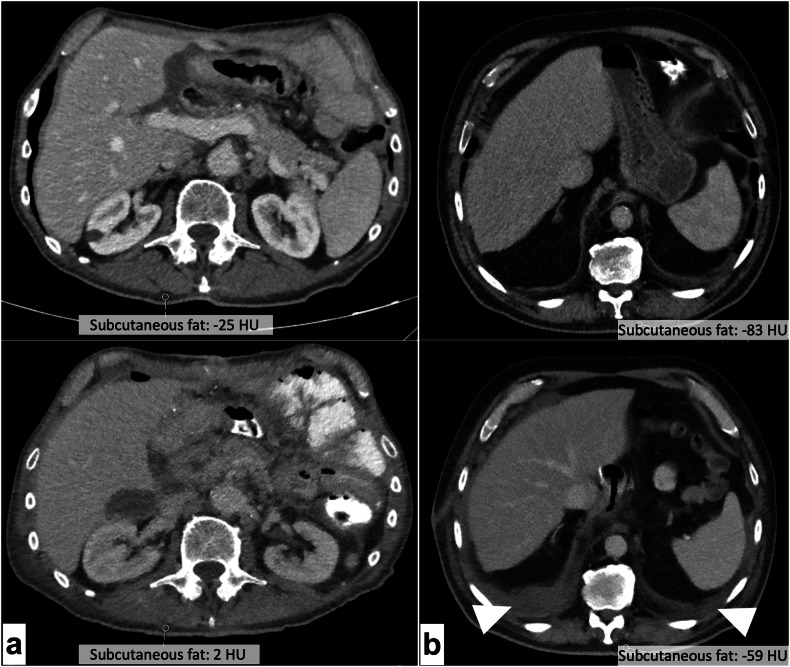
Fig. 5.
a Axial reformations of two scans of the same patient, obtained 16 days apart at the L2 level (top: baseline; bottom: follow-up). Derived bone mineral density measurements changed significantly between scans (baseline: 148.3 mg CaHA/cm3; follow-up: 175.1 mg CaHA/cm3). Manual case review revealed that the patient suffered multiple intraabdominal abscesses between scans. Subcutaneous fat HU increased from -25 to +2 between scans as sign of hydropic decompensation. Also note the progressive mesenterial fluid injections and paracolic ascites. b Imaging at the L1 level revealed pleural effusion in the follow-up scan (bottom) of this patient, 19 days after baseline (top). The patient also showed an increase of subcutaneous fat attenuation from -83 HU at baseline to -59 HU at follow-up, co-occurring with an increase in measured bone mineral density (baseline 111.4 mg CaHA/cm3; follow-up: 133.1 mg CaHA/cm3)

