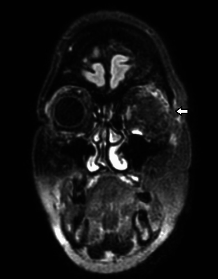
Figure 1. Pre-operative MRI FLAIR scan.
Meningioma of the left sphenoid wing, with projection into the temporal fossa and orbit, with diffuse meningeal extension to the temporal squama and left frontal bone. It reduces the left orbit, which compresses the muscles and optic nerve in the posterior part and causes proptosis.

