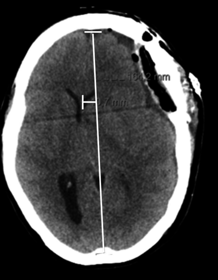
Figure 3. Axial plane of brain computed tomography at the third post-operative day and 24 hours under sedation.
A pericerebral hypodense collection remains, currently extending to the high fronto-parietal convexity. Consequently, there is a greater deformation of the brain parenchyma, resulting in a deviation of the midline to the right, currently approximately 11 mm, a degree greater than that shown in the previous examination (Figure 2).

