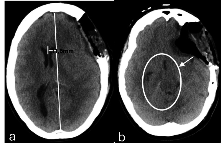
Figure 4. Axial plane of brain computed tomography after decompressive craniectomy.
(a) Mass effect remains on the left cerebral hemisphere and on the ventricular system, with signs of subfalcine hernia and clear deviation of the midline structures to the right, approximately 11.5 mm (the previous examination was approximately 10.7 mm). (b) An outline of an uncal hernia (white arrow), with molding of the midbrain and decreased permeability of the base cisterns (white circle).

