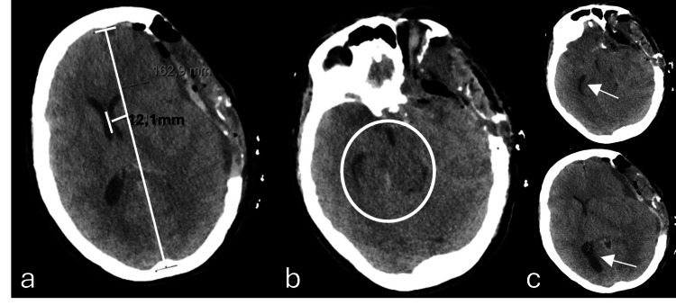
Figure 5. Axial plane of brain computed tomography taken 12 hours after post-craniectomy enlargement in the left temporoparietal region (third surgical intervention) due to neurological deterioration with fixed mydriasis.
There is evidence of bi-hemispheric convexity grooves effacement, base cisterns effacement, and 12 mm midline deviation to the right (a), as well as subfalcine herniation and inferior transtentorial herniation of the left uncus, with marked deformation of the midbrain (b). There is a slight increase in the amplitude of the temporal horn (superior arrow) and the occipital horn (inferior arrow) of the right lateral ventricle, reflecting an increase in internal CSF tension, due to disturbance of normal circulation (c).

