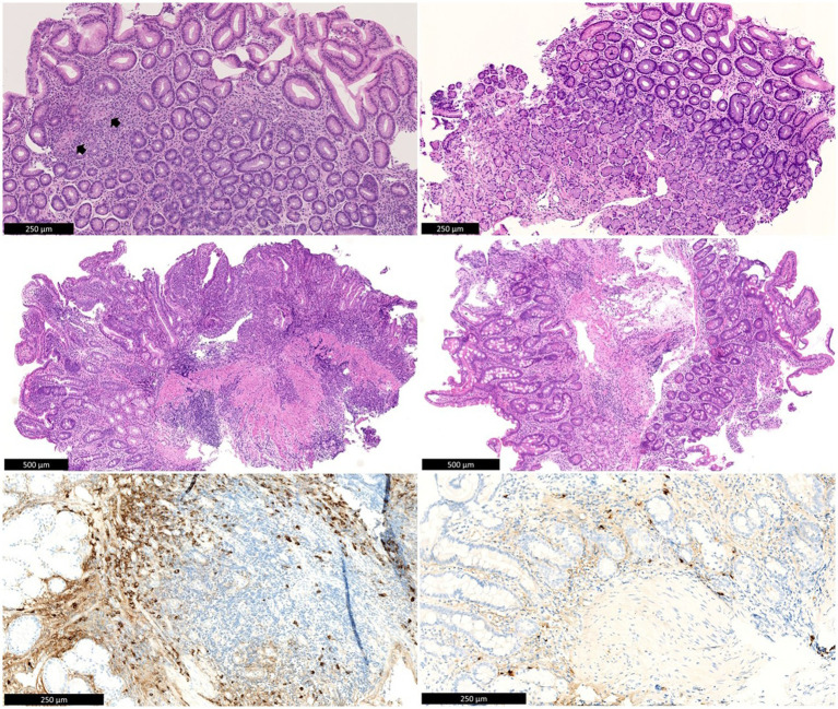
Figure 1.
Top left: Antral mucosa with chronic inactive gastritis and two non-necrotic granulomas (arrows). Middle left: Duodenal bulb mucosa with active chronic inflammation with erosion. Below left: IgG4 immunohistochemistry shows numerous IgG4-positive plasma cells in the duodenal mucosa (>100/HPF). There is background staining in the stroma, as is often seen with high serum IgG4 levels. Top right: Antral mucosa after treatment, without significant inflammation. Middle right: Duodenal mucosa after treatment, without significant inflammation. Below left: IgG4 immunohistochemistry shows rare residual IgG4-positive plasma cells in the duodenal mucosa. There is also much weaker background staining in the stroma compared to before treatment.

