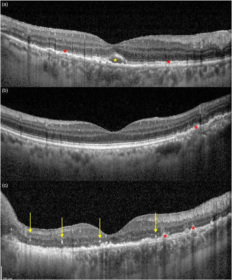
Figure 3.
B-scan Optical coherence tomography (OCT) of three biopsy-proven VRL. Retinal pigment epithelium (RPE) thickening and mottling can be disclosed, as well as small sub-RPE deposits (a, b and c, red asterisks). A small macular subretinal hypo-reflective deposit is marked in A (yellow asterisk). Vertical hyper-reflective spots are signs of intraretinal lymphomatous infiltration and have been highlighted in C (yellow arrows).

