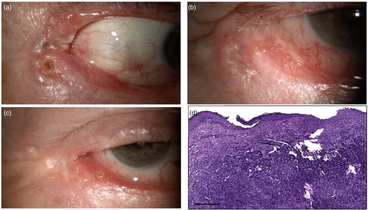
Figure 4.
(a) Sclerotic, ulcerated lesion infiltrating the lower eyelid and extending to the lateral canthus and toward the upper eyelid; (b) Lateral canthus after surgical excision; (c) Lateral canthus 2 weeks after IRT; (d) Basal cell carcinoma, nodular type: Histopathological examination showed neoplastic islands of basaloid neoplastic cells with hyperchromatic nuclei and scant cytoplasm, with peripheral artifactual clefting (hematoxylin and eosin stain, original magnification x10, scale bar 100 µm).

