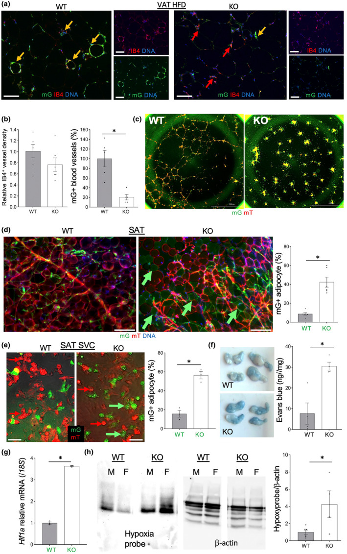
FIGURE 2.

Tert KO causes endothelial cells (EC) dysfunction, mis‐differentiation, and vessel leakiness. (a) Anti‐GFP IF analysis in tissue sections, with IB4 counterstaining EC, reveals a lack of mG+ cells in blood vessels in VAT of EC‐Tert‐KO mice fed HFD (9 months). (b) Data quantification from (a) based on IB4 binding and mG expression in the vasculature. (c) Self‐organization of SAT mG+/mT+ SVC into stromal‐vascular networks showing a defect of Tert‐KO EC. (d) Whole mount of SAT showing increased frequency of EC‐derived adipocytes in AT of EC‐Tert‐KO mice. Graph: Data quantification. (e) Primary culture of SVC from SAT after adipogenesis induction showing increased differentiation of Tert‐KO mG+ cells into adipocytes containing lipid droplets. Graph: Data quantification. (f) VAT 0.5 h after intravenous injection of Evans blue and subsequent systemic perfusion showing increased dye retention in EC‐Tert‐KO mice. Graph: Dye retention quantification. (g) q‐RT‐PCR reveals higher Hif1a expression (normalized to 18S RNA) in mG+ cells FACS‐sorted from SAT of EC‐Tert‐KO mice (8 months old). (h) Extracts from VAT 0.5 h after injection of hypoxyprobe analyzed by PAGE. β‐Actin immunoblotting: Loading control. Note increased hypoxyprobe retention in VAT of EC‐Tert‐KO males (M) and females (F). Graph: Data quantification: Mean ± SEM. For all data, shown are mean ± SEM (error bars). *p < 0.05 (two‐sided Student's t‐test). Scale bar = 50 μm.
