FIGURE 4.

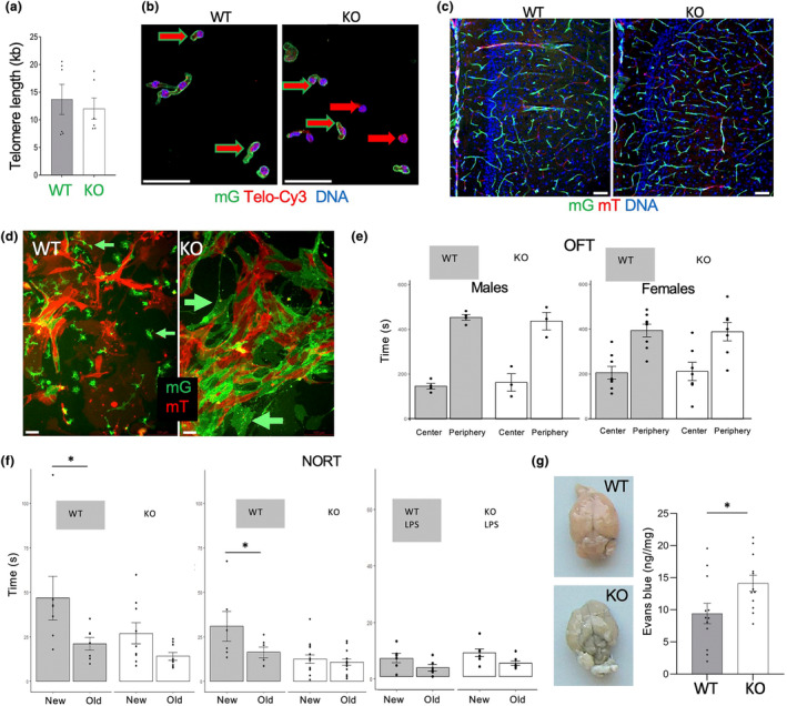
Tert KO impairs brain ECs and cognitive function without telomere attrition. (a) q‐PCR on DNA from brain mG+ lineage cells of EC‐Tert‐KO mice at 10 months of age. Real‐time PCR data are normalized to data for a single copy gene. (b) Telo‐FISH reveals comparable telomere length (red TelC‐Cy3 signal) in mG+ cells (green outline arrow) from EC‐Tert‐KO and WT mice. (c) mG and mT fluorescence in hypothalamus sections reveals normal vasculature in EC‐Tert‐KO mice. (d) Primary culture of cells from the brains 2 days after plating at identical density. Note reduced proliferation and larger size of mG+ EC‐Tert‐KO cells. (e) Open field test does not reveal behavioral abnormality in EC‐Tert‐KO mice. (f) Novel object recognition test (NORT) reveals memory impairment in EC‐Tert‐KO males and females. Lipopolysaccharide (LPS) injection, impairing memory, was used as a positive control. (g) Brains 0.5 h after tail vein injection of Evans blue and subsequent systemic perfusion showing increased dye retention in EC‐Tert‐KO mice. Graph: calorimetric Evans blue quantification. For all data, shown are mean ± SEM (error bars). *p < 0.05 (two‐sided Student's t‐test). Scale bar = 50 μm.
