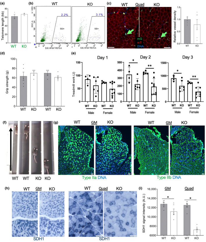
FIGURE 5.

Tert KO impairs muscle ECs and function without telomere attrition. (a) q‐PCR on DNA from combined quadriceps (Quad) and gastrocnemius (GM) muscle mG+ lineage cells of EC‐Tert‐KO mice at 10 months of age. Real‐time PCR data are normalized to data for a single copy gene. (b) Flow cytometry on hindlimb skeletal muscle cells recovered 3 days after EdU injection comparing incorporation into mG+ and mT+ cells. (c) Fluorescence analysis of mG+ and mT+ cells in Quad muscle. Graph: data quantification based on vascular mG expression. (d) Grip strength, measured for forelimb and hindlimb of EC‐Tert‐KO and WT littermates (N = 5). (e) Quantification of data from e for 10 daily runs of N = 5 WT and N = 5 EC‐Tert‐KO mice, showing reduced physical endurance of EC‐Tert‐KO mice, reflected in Joules of work performed (weight (kg) × speed (m/min) × time (min) × incline (degree) × 9.8 m/s2). (f) A snapshot of WT and EC‐Tert‐KO littermates running on a treadmill (arrows: direction) illustrating reduced fatigue resistance capacity in EC‐Tert‐KO mice. (g) IF analysis of muscle fiber types in GM muscle. (h) Immunohistochemistry on GM and Quad reveals decreased succinate dehydrogenase (SDH1) expression in the muscle fibers of EC‐Tert‐KO mice. (i) SDH1 expression quantification from (h). For all data, shown are mean ± SEM (error bars). *p < 0.05 (two‐sided Student's t‐test). Scale bar = 50 μm.
