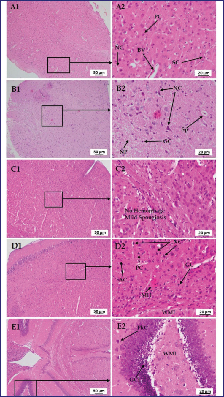
Figure 11. Photomicrograph of histo-structures of brain of different treatment groups.Group A-Control, Group B-LPS treated group, Group C-LPS and Polymyxin B treated group, Group D-LPS and honey treated group, Group E-LPS, Polymyxin B and honey treated group. Control group A showed normal tissue structures with regularly organized neuron cell (NC), pyramidal cell (PC), satellite cell (SC), and blood vessel (BV) in the cortical layer of the cerebrum.But in the case of LPS-treated group B, several nerve fiber degenerations, and spongiosis (Sp) were found. Whereas, neuropil (NP) and glial cells (GC) were normal. In the case of group C, no hemorrhage but mild spongiosis (Sp)wasfound. In the case of group D, most of the cells are normal along with astrocytes (AC) and white matter layer (WML) except mild hemorrhage (MH) and vacuolation. However, in group E, there were no detectable changes in the granular cell layer (GCL), white matter layer (WL) and Purkinje cell (PkC) were also normal.

