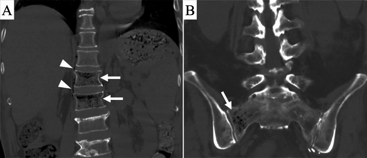
Figure 1. Computed tomography (CT) images of the lumbosacral spine obtained 112 days after liver transplantation.
(A) Coronal CT image of the lumbar spine shows clusters of small intraosseous gas (arrows) in the first and second lumbar vertebral bodies, extraosseous gas (arrowheads) around the vertebral bodies, and compression fractures of the first and second lumbar vertebral bodies.
(B) Coronal CT image of the sacrum shows clusters of small intraosseous gas (arrow) in the right sacral ala.

