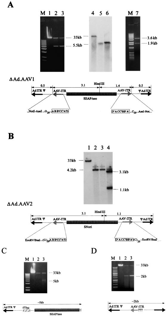
FIG. 2.
Production and characterization of Ad-AAV hybrid vectors. (A) Ad.AAV1 was amplified in 293 cells and banded in a CsCl step gradient. Undigested viral DNA purified from CsCl gradient fractions was analyzed in 0.8% agarose gels: lane 1, DNA isolated from 10 μl of banded Ad.AAV1 particles (full-length, 35-kb genome); lanes 2 and 3, DNA isolated from 10 μl of the banded ΔAd.AAV1 particles (5.5-kb genome) after one CsCl step gradient (from two different virus preparations). Viral material containing ΔAd.AAV1 genomes was purified by additional CsCl equilibrium gradients. Viral DNA (100 ng) from purified particles was analyzed by Southern blotting with a SEAP-specific probe. Lane 4, full-length Ad.AAV1 DNA; lanes 5 and 6, ΔAd.AAV1 particles purified by one (lane 5) or two (lane 6) rounds of ultracentrifugation in equilibrium gradients. Viral DNA (1 μg) from the 5.5-kb product of Ad.AAV1 isolated from purified particles was digested with HindIII and was analyzed in a 0.8% agarose gel (lane 7). The structure of the 5.5-kb genome (ΔAd.AAV1) shown at the bottom of panel A was deduced after restriction and sequence analysis. The position of the HindIII site is indicated. The AAV ITR-vector junctions were sequenced with primers specific to regions within the transgene cassette (see Materials and Methods). The palindromic composition of the AAV ITRs is framed. M, 1-kb DNA ladder (GIBCO-BRL). (B) Ad.AAV2 was amplified and CsCl banded in the same way as described for Ad.AAV1. Viral material was analyzed as for Ad.AAV1 (lanes 1 to 3). The amount of DNA per lane corresponds to 5 μl of viral material. The faint bands that run above Ad.AAV2 (lane 1) and ΔAd.AAV2 DNA (lanes 2 and 3) may represent a different conformational structure, which was seen often with linear plasmid DNA containing AAV ITRs. Lane 4; HindIII digest of ΔAd.AAV2 DNA. (C and D) Amplification products of Ad.AAV-GC and Ad.AAV1-Δ1ITR, respectively. Viral DNA in lanes 1 was isolated from 5-μl samples of banded viruses with full-length genomes. Viral DNA separated in lanes 2 and 3 was isolated from 1-ml gradient fractions with densities lighter than 1.32 g/cm3. No banded virus was visible in these fractions.

