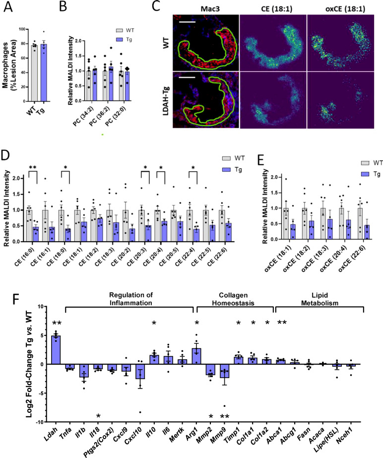
Fig. 7. LDAH impacts sterol homeostasis in vivo and leads lesional foam cells to a less inflammatory phenotype with a pro-fibrotic molecular signature.
A–E Atherosclerotic lesions of 20-week-old LDAH-Tg (Tg, blue bars) (n = 5) and wild-type (WT, gray bars) (n = 6) littermate male mice were analyzed using high-resolution MALDI-MSI. A Macrophage content was similar in lesions of mice of both genotypes. B Quantification of main structural PC species by lesion area using MALDI-MSI. C Exemplary images of consecutive sections stained for Mac3 and MS images representing CE (18:1) and oxidized (ox) CE (18:1). Scale bars: 200 μm. (D, E) Quantification of main CE (D) and candidate oxidized CE (E) species identified in atherosclerotic lesions. The first number in the parenthesis represents the number of carbons of the fatty acid esterified to the sterol, and the number after the colon represents the number of double bonds. F Gene expression analyzes. RNA was obtained by LCM from Mac3 positive areas within lesions of LDAH-Tg (Tg) and wild-type (WT) male mice (n = 5 per genotype). The blue bars represent Log2-fold change of Tg over WT. Comparisons were performed by two-tailed unpaired t-test (normally distributed with equal variances), two-tailed Welch’s t-test (normally distributed with unequal variances), or two tailed Mann-Whitney U (not normally distributed). All data in this figure are presented as mean ± SEM of independent samples. *P < 0.05, **P < 0.01, ***P < 0.001. Source data are provided as a Source Data file.

