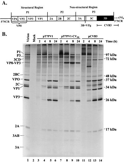
FIG. 1.
Capsid processing of PV1 chimeras containing CVB3 3Dpol. (A) Schematic representation of a full-length chimeric genome and viral polyprotein organization are shown. The viral structural proteins are derived from the P1 portion of the genome, and the nonstructural proteins are derived from the P2 and P3 portions of the genome. PV sequences are represented by solid lines and white boxes, whereas CV sequences are represented by the black box (3D coding sequences) and dotted lines (3′ NCR). (B) In vitro translation of PV1, CVB3, and chimeric T7 transcripts. Full-length transcripts from wild-type PV1 cDNA (lanes 3 to 6), chimeric PV1-CVB3 3D cDNA (lanes 7 to 10), and wild-type CVB3 cDNA (lanes 11 to 14) were used to program translation reactions in HeLa S10-ribosomal salt wash extracts. At the indicated times, aliquots were removed, diluted in Laemmli sample buffer, and subjected to electrophoresis on a 12.5% polyacrylamide–SDS gel. Lane 2 contains a mock translation mixture incubated for 8 h with no RNA added. A translation reaction mixture programmed with PV1 virion RNA was loaded in lane 1 as a marker for viral proteins. The positions of viral proteins are indicated to the left and the molecular masses are indicated to the right.

