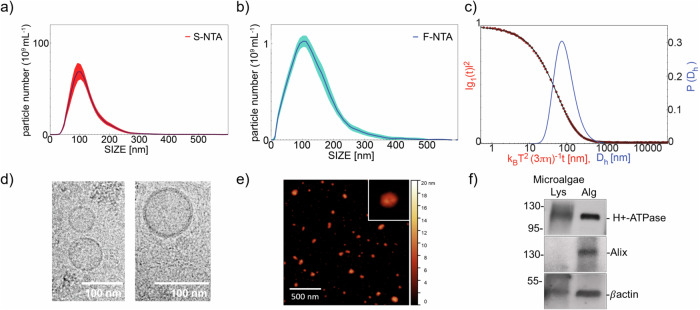
Fig. 1. Quality control of nanoalgosome preparations.
a Nanoalgosome size distribution and concentration measured by NTA, the red deviation is relative to the five measurements analysed per sample. b Size distribution of nanoalgosomes stained with Di-8-ANEPPS measured by F-NTA (using laser wavelength of 488 nm), the blue deviation is relative to five measurements analysed per sample, and (c) DLS. Morphology of nanoalgosomes was analyzed by (d) cryo-TEM and (e) AFM. f Immunoblot analyses were performed on T.chuii lysate (Lys) and nanoalgosomes (Alg) to detect the markers H+-ATPase, Alix and β-actin. Representative results of three independent biological replicates (n = 3 biologically independent samples) are presented.

