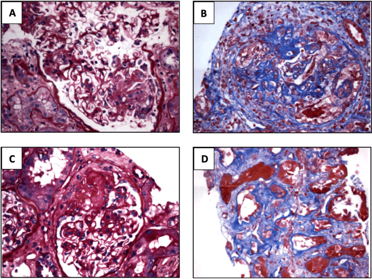
Figure 3. Kidney biopsy sections stained with H&E, PAS, trichrome, and JMS (magnification 20x).
A. Cellular crescent B. Fibrinoid necrosis C. Fibrous crescent D. Interstitial fibrosis
Kidney biopsy demonstrated focal necrotizing and diffuse crescentic glomerulonephritis, pauci-immune type with mild activity and moderate chronicity. Tubular atrophy and interstitial fibrosis were moderate

