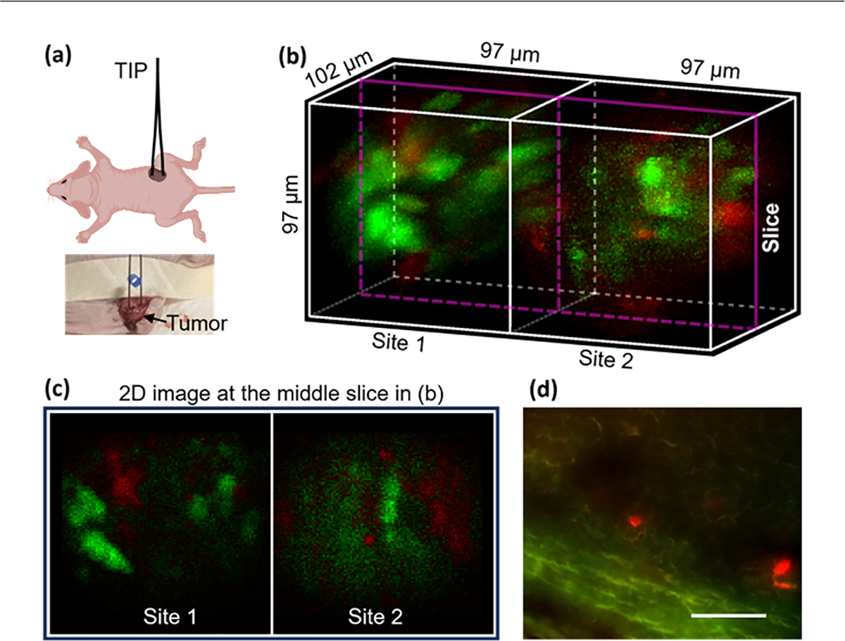
Fig. 5.

Two-site in vivo imaging of a hybrid tumor in a living mouse. (a) Schematic and photo of the testing in the animal, (b) an acquired image (green: eYFP; red: mCherry), (c) 2D image at the middle slice in (b), and (d) image of a cryosection of the tumor, scale bar: 60 μm.
