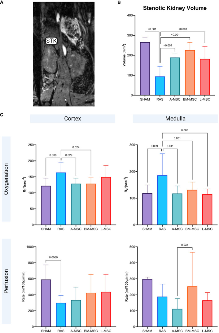
Figure 2.
Representative MRI image of STK in coronal section (A). Non-invasive measurement of volume in the STKs (B) and the oxygenation and perfusion to the cortex and medulla in the STKs (C) within each group. All measurements are expressed as mean ± SD. For oxygenation, R2*(sec-1) reflects hypoxia with lower R2* indicating better oxygenation. STK, stenotic kidney.

