FIG. 3.
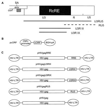
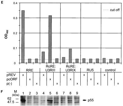
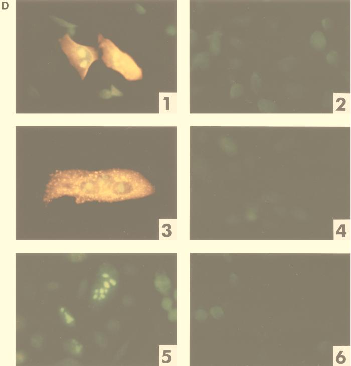
Mapping of the HTDV/HERV-K cORF-responsive element. (A) Mapping of RcRE: genomic localization of 3′-LTR subfragments tested for RRE-like activity in combination with cORF. Fragments depicted as solid lines were active; that depicted by a hatched line was inactive. (B) Mammalian expression vector encoding HTDV/HERV-K cORF (effector plasmid). BGH, bovine growth hormone; CMV, cytomegalovirus; pA, polyadenylation sequence. (C) Mammalian HIV Gag expression vectors containing the HIV RRE or different sections of the HTDV/HERV-K 3′ LTR (U3RIII, nt 8720 to 9149; U3RIX, nt 8720 to 9093; RU5, nt 9065 to 9492) and the negative-control vector lacking an RRE element (reporter plasmid). (D) Cotransfection of reporter and effector plasmids into HLtat cells. Panel 1, pHIVgagU3RIII and pcORF; panel 2, pHIVgagU3RIII and p(−); panel 3, pHIVgagU3RIX and pcORF; panel 4, pHIVgagU3RIX and p(−); panel 5, pHIVgagRU5 and pcORF; panel 6, pHIVgagRU5 and p(−). Staining was performed with a mouse monoclonal α-HIV p24 antibody (panels 1 to 6) (orange) and with a rabbit α-cORF antibody (panels 1, 3, and 5) (green). A filter suitable for both fluorescence markers was used. (E) Determination of the HIV Gag precursor levels in particle preparations by an HIV p24 capture assay (Abbott). HLtat cells were transfected with the vector combinations listed in the table below. The cutoff value was determined by the extinction of the lysis buffer. (F) Detection of the HIV Gag precursor by Western blot analysis. The p55 Gag precursor was present in combination 1 (Rev and RRE), in 5 and 8 (cORF and RcRE), and faintly in 4 and 7 (Rev and RcRE). The positions of molecular mass markers (M) are indicated on the left.


