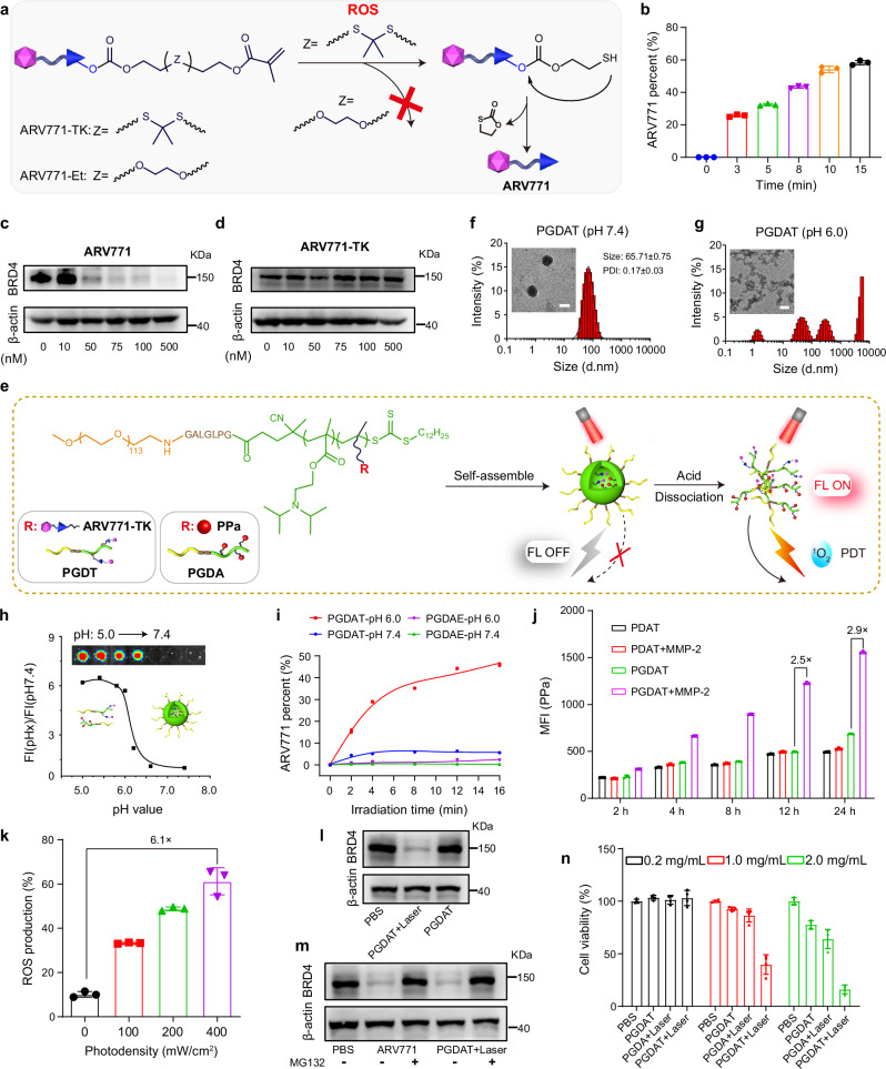
Fig. 2. Synthesis and characterization of the ROS-activatable PROTAC nanoparticle.
a Schematic illustration of ROS-induced restoration of ARV771 from ARV771-TK, but powerless of the counterpart ARV771-Et. b Release rate of ARV771 from ROS-activatable ARV771-TK after being mixed with PPa and irradiated by 671 nm laser with determined time (n = 3 independent experimental units). c, d Western blot analysis of ARV771 and ARV771-TK mediated BRD4 degradation in MDA-MB-231 cells post 24 h of co-incubation. e Schematic diagram of the self-assembly process and acid-activatable photoactivity capability of PGDAT nanoparticle. Representative DLS data and TEM images of the PGDAT nanoparticle at pH 7.4 (f) and pH 6.0 (g) condition (scale bar = 50 μm). h Acid-induced fluorescence recover profile of PGDAT nanoparticle, the fluorescence intensity was normalized to that determined at pH 7.4. Insert image was the PGDAT nanoparticle solutions at different pH values. i ROS-triggered ARV771 release from the PROTAC nanoparticles at the pH 7.4 and 6.0 with the different laser exposure time (n = 3 independent experimental units). j Flow cytometry evaluation of the cellular uptake of MMP-2-responsive PGDAT nanoparticle and MMP-2-inert PDAT nanoparticle by MDA-MB-231 cells after co-incubation with defined time in vitro (the nanoparticles were pretreated with/without MMP-2 of 0.2 mg/mL for 1 h) (n = 3 independent experimental cell lines). k Flow cytometric analysis of ROS production activity of PGDAT nanoparticle, DCFH-DA probe was joined into the tumor cells before laser irradiation with different photodensity (n = 3 independent experimental cell lines). l Western blot examination of BRD4 degradation of ROS-activatable PGDAT nanoparticle after 24 h co-incubation with MDA-MB-231 cell, the PGDAT nanoparticle was pretreated with 671 nm irradiation or not. m Western blot assay of MDA-MB-231 cells which were subjected to ARV771 and PGDAT nanoparticle with or without MG132 treatment (identical PROTAC concentration of 1.0 μM and MG132 concentration of 5.0 mM). n CCK-8 assay of MDA-MB-231 cell viability post different treatments (n = 3 independent experimental cell lines). All data are presented as mean ± SD.

