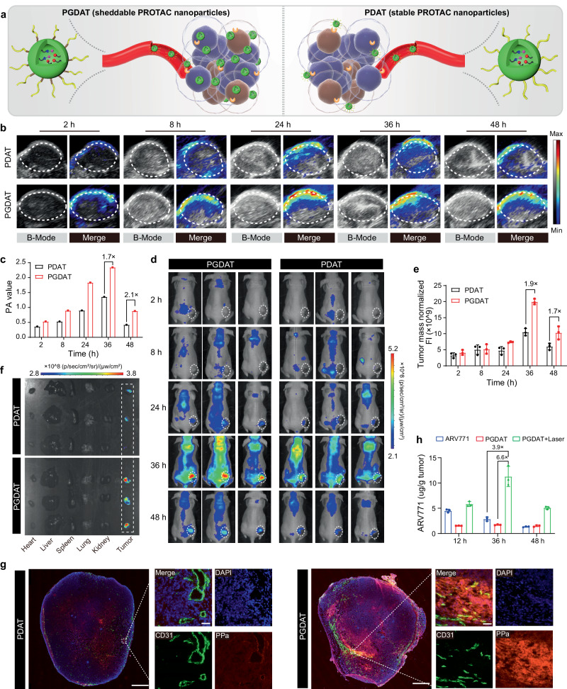
Fig. 3. Stimuli-activatable PROTAC nanoparticle specifically accumulated and released free PROTAC at the tumor site in vivo.
a Schematic diagram of sheddable PROTAC nanoparticles integrating MMP-2-lible GPLGLAG peptide spacer for enhanced tumor accumulation and penetration compared to the MMP-2-insensitive counterpart (without GPLGLAG peptide spacer). b Photoacoustic images (PAI) of PROTAC nanoparticle distribution in MDA-MB-231 tumor-bearing mouse model in vivo. c PA value of the tumor site (n = 3 mice). d Fluorescence imaging analysis of PGDAT (with MMP-2 responsive spacer) and PDAT (MMP-2-insensitive) nanoparticles distribution in MDA-MB-231 tumor-bearing nude mice. e Normalized fluorescence intensity of tumor site (n = 3 mice). f Ex-vivo fluorescence images of the harvested major organs (heart, liver, spleen, lung, kidney) and tumor tissues post 48 h treatment. g Ex-vivo CLSM images of tumor section post 48 h injection (left panel scale bar = 2.0 mm, right panel scale bar = 50 μm, the blue represents DAPI, the green represents CD31 and the red represents PPa). h HPLC evaluation of the intratumoral ARV771 distribution with different treatments (ARV771, PGDAT and PGDAT + laser groups with the identified ARV771 dose of 10 mg/kg) (n = 3 mice). All data are presented as mean ± SD.

