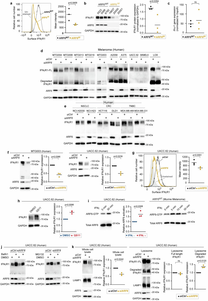
Fig. 6. ARF6-dependent IFNγR1 surface expression in murine and human melanoma.
a Flow cytometric detection of IFNγR1 cell surface expression in early-passage murine tumour cell lines, n = 3 biologically independent tumour cell lines of each genotype. b Western blot for IFNγR1 in early-passage murine tumour cell lines, n = 3 biologically independent tumour cell lines of each genotype. c Quantitative RT-PCR for Ifngr1 in n = 3 biologically independent, early-passage murine tumour cell lines of each genotype. d, e Western blot for full length (FL) and degraded IFNγR1, ARF6 and GAPDH in human melanoma patient-derived xenograft cells (MTG) and commercially available melanoma lines (d), in human non-small cell lung cancer (NSCLC), colorectal cancer (CRC), triple negative breast cancer (TNBC) cell lines (e). f Quantification of IFNγR1 in MTG003 (n = 3 biologically independent experiments) and UACC.62 (n = 3 biologically independent experiments) with or without ARF6 knockdown. g Flow cytometric detection of surface expression of IFNγR1 in UACC.62 with or without ARF6 knockdown, n = 3 biologically independent experiments. h Western blot for IFNγR1 in UACC.62 without or with 2 µM QS11 treatment for 24 h, n = 3 biologically independent experiments. i Western blot for total ARF6 and ARF6-GTP in UACC.62 human melanoma cells and murine melanoma cells with or without 500 U/mL IFNγ treatment, n = 3 biologically independent experiments. j Western blot for indicated proteins in UACC.62 with or without ARF6 knockdown and with or without 50 nM Bafilomycin A1 or 10 µM MG132 treatment for 6 h. n = 1 experiment. k Western blot analyses of UACC.62 with or without ARF6 knockdown as indicated, n = 3 biologically independent experiments. a–c, g Two-tailed t-test. f, h, i, k Two-tailed Ratio paired t-test. a–c, f–h, i, k Solid line within data points = mean. See also Supplementary Fig. 6. Source data are provided as a Source Data file.

