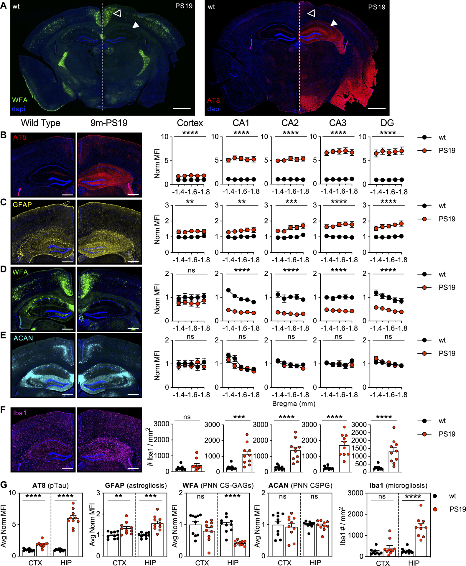
Figure 1. 9m-PS19 mice exhibit loss of WFA+ PNNs in the presence of pTau accumulation and gliosis.

9-month-old PS19 mice exhibit A) striking loss of hippocampal WFA+ PNNs in association with regional pTau accumulation. 5-region stereology analysis (Bregma −1.4 to −1.8 mm) of B) AT8 (pTau), C) GFAP (astrogliosis), D) WFA (PNN CS-GAGs), E) ACAN (PNN CSPG), and F) Iba1 (microgliosis) were performed and then G) averaged and quantified. Solid arrows: dorsal hippocampus; Open arrow: retrosplenial cortex. Scale bar: A) 1 mm, B-F) 0.5 mm, representative images from male mice, dapi included in all images. Statistics reported in Supplemental Table 1.
