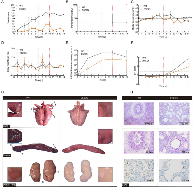
Figure 5.
CD163-E529G mutation pigs were resistant to HP-PRRSV infection
A–D: Relative clinical symptoms (A), survival rate (B), rectal temperature (C), and body weight gain rate (D) in challenged WT (black line) and E529G (brown line) pigs were monitored daily. Survival rate showed that two WT pigs died at 7 and 10 days, respectively (red dashed line). E, F: Blood samples from challenged pigs were collected on days 0, 3, 7, 10, and 14. Viral load (E) and viral antibody levels (F) in serum were separately determined by qPCR and ELISA. G: Each photo is from a single representative pig. Macroscopic observation of lung (above), spleen (medium), and lymph node (below) from WT (left) and E529G (right) pigs, and locations of tissue lesions are marked with black arrows, partial lesion locations are enlarged. H: Histopathological diagnosis in lungs between WT (left) and E529G (right) pigs was indicated by H&E staining, with 10× magnification shown above and 20× magnification shown in the middle. Viral infection in lungs from WT and E535G pigs was determined by IHC (below).

