
Fig. 1.
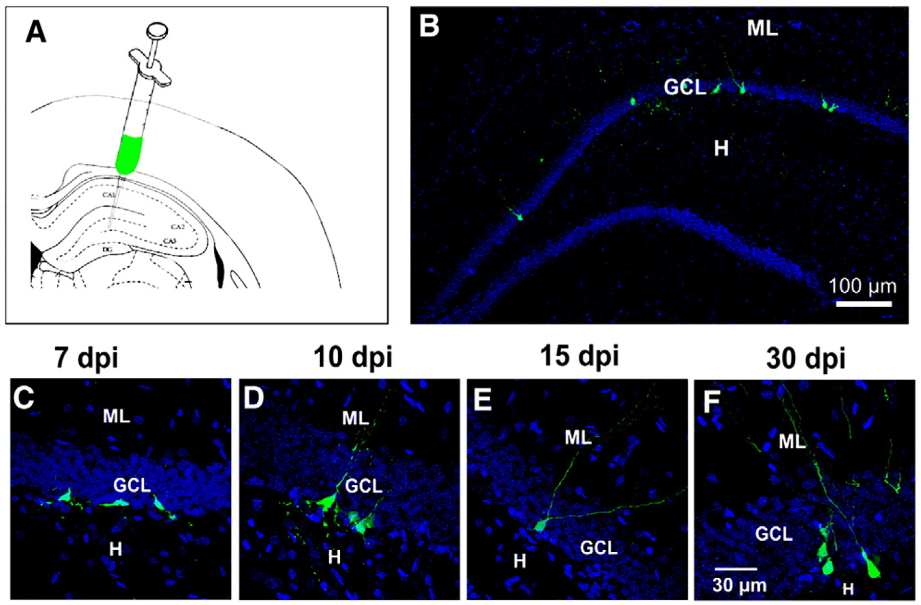
Newborn cells were detected in adult rats by retroviral infection. A) Schematic view of the hippocampus depicting the site of injection of the retrovirus. B) Panoramic view of a dentate gyrus with GFP+ cells in the inner granule cell layer at 15 dpi. Confocal images showing the morphology of GFP+ cells at the post-injection times analyzed: C) at 7, D) at 10, E) at 15 and F) at 30 dpi. DAPI is shown in blue. ML, molecular layer; GCL, granule cell layer; H, hilus. Calibration in F applies to panels C–E.