
Fig. 5.
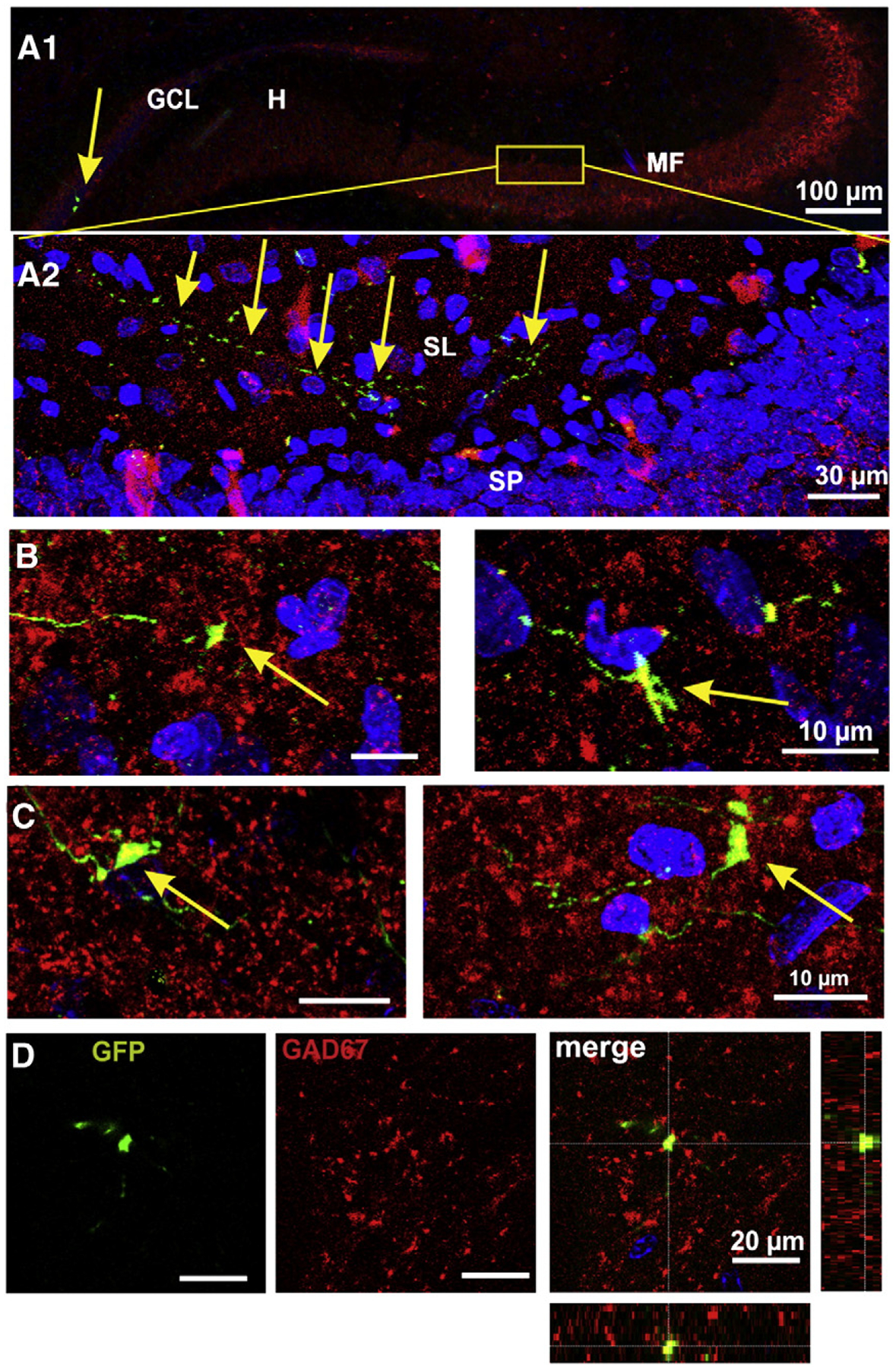
The mossy fibers express GAD67. A1) Panoramic view of the mossy fiber projection showing, on the left hand, two GFP+ GCs (arrow) and mossy fibers. The area shown at high magnification in A2 is signaled. A2) Confocal image of an axon of a GFP+ granule cell is shown along the stratum lucidum of CA3. B) Confocal images (50 optical slices of 0.5 μm) of mossy fiber giant boutons expressing GAD67 at 15 dpi. C) Mossy fiber boutons of GFP+ GCs at 30 dpi, did express GAD67 enzyme. D) Three-dimensional reconstructions through the Z-axis of GFP+ boutons (fifty optical slices at 0.5 μm intervals) were used to verify the co-localization of GFP and GAD67. Orthogonal projections are shown on the right most panel.