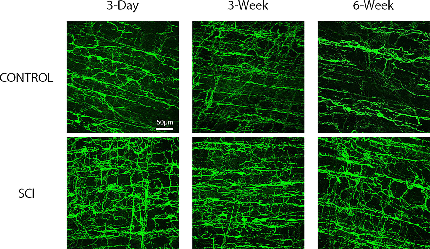
FIGURE 2.

Representative male rat surgical control and spinal cord injured (SCI) maximum projection confocal images of whole mounted distal colon processed for c-Kit immunofluorescent identification of myenteric and smooth muscle Interstitial Cells of Cajal (ICC). c-Kit immunofluorescence demonstrates an increase in pixel density beginning 3-days post-SCI compared to corresponding surgical controls, and this pattern is maintained at the 3 and 6- weeks time points. Images are 40× with scale bar = 50 μm.
