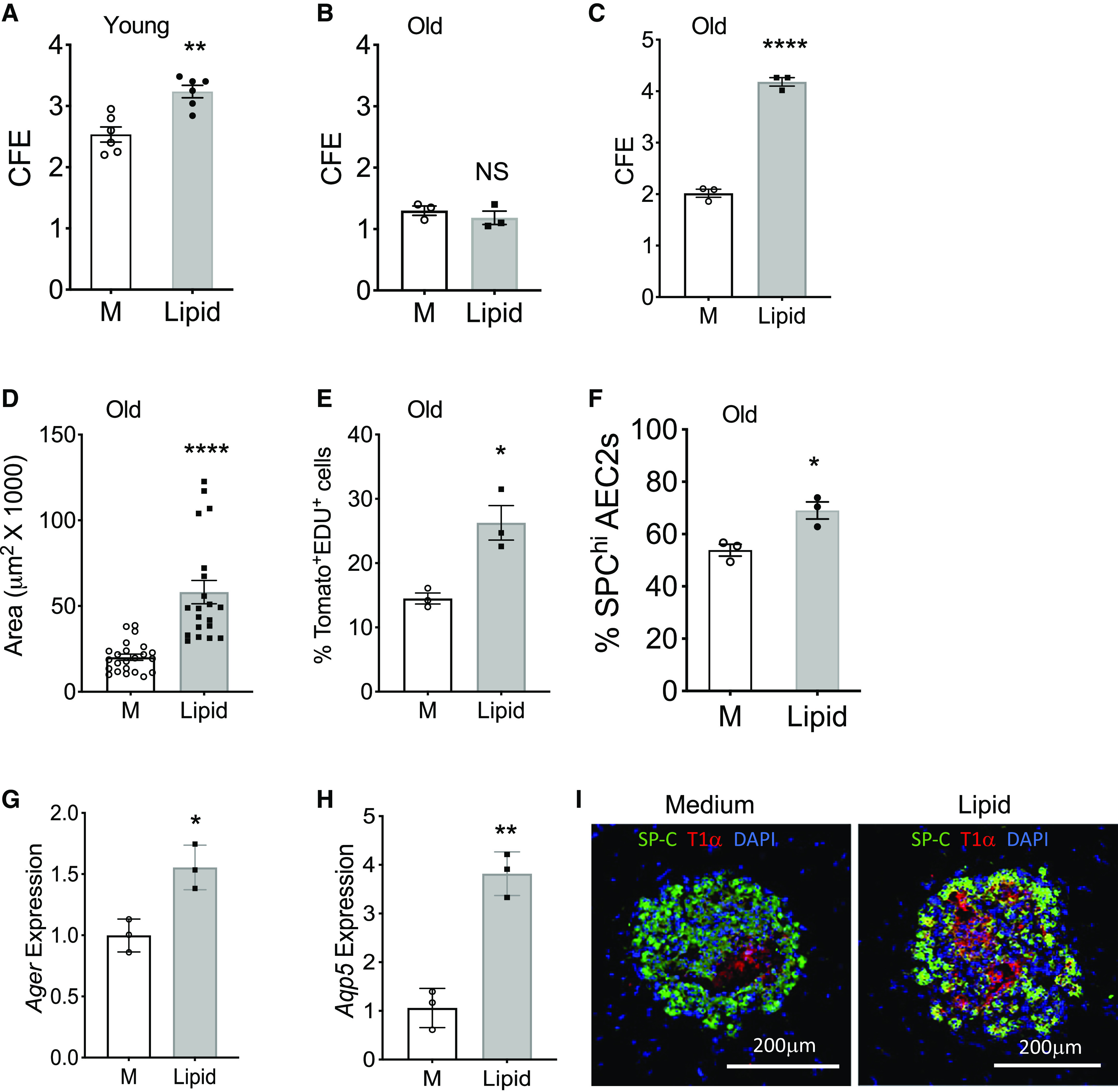
Figure 7.

Lipid supplementation promoted renewal capacity of mouse AEC2s. (A and B) CFE of AEC2s from young (A; n = 6; **P < 0.01) and old mice (B; n = 3) with and without 2% lipid treatment. M = medium control. (C–E) 3D organoid cultures of AEC2s from 20-month-old tamoxifen-treated SFTPC-CreER+ Rosa-Tomatofl/fl mice with and without 4% lipid treatment. CFE (C; n = 3; ****P < 0.0001); Sizes of colonies (D; n = 20–23; ****P < 0.0001), and the percentage of EdU+ AEC2s in gated total Tomato+ AEC2s derived from 3D cultured organoids by flow cytometry (E; n = 3; *P < 0.05). (F) Freshly isolated lung single cells from 24-month-old mice were cultured with and without lipid supplementation for 48 hours. The percentage of SPChi cells in total gated AEC2s was determined by flow cytometry (n = 3; *P < 0.05). (G and H) Expression levels of Ager and Aqp5 in young mouse AEC2s after 3D culture with and without 2% lipid were assessed with RT-PCR (n = 3; **P < 0.01 and *P < 0.05). P values were calculated by unpaired two-tailed Student’s t test. (I) Representative images of 3D organoids derived from young mouse AEC2s and stained with SFTPC and T1α antibodies (n = 6). Scale bars, 200 μm.
