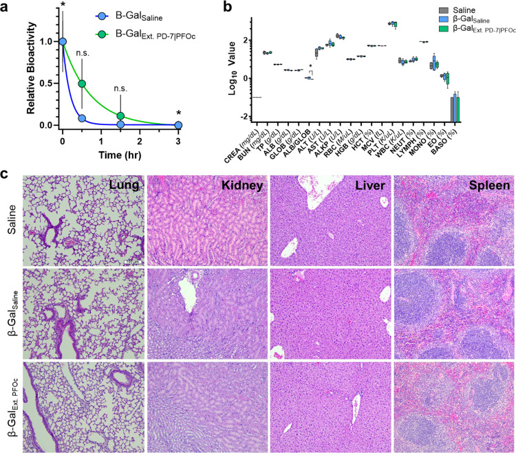
Figure 6.
In vivo half-life and safety of fluorous proteins. (a) Time-dependent activity of β-Gal in plasma delivered intravenously in either saline (β-GalSaline, blue) or extracted PFOc (β-GalExt. PFOc, green). Data shown as average ± s.d. of n = 5 technical replicates. Statistical significance calculated with Student’s t test, with n.s. = not significant and *p < 0.05. (b) Serum toxicology results from C57BL/6J mice 24 h after injection of saline (control), β-GalSaline, or β-GalExt. PFOc. Data shown as box and whisker plot ± s.d. of n = 5 technical replicates. Statistical significance calculated using Student’s t test and represented as * p < 0.05; data subsets with p > 0.05 represented nonsignificant changes. (c) Representative histology images of lung, kidney, liver, and spleen tissue section from C57BL/6J mice 24 h after administration of saline (control), β-GalSaline, or β-GalExt. PFOc. Each imaging group consisted of n = 4 mice, with detailed tissue section evaluation conducted by an unblinded board-certified veterinary anatomic pathologist. Scale bar: 100 μm.

