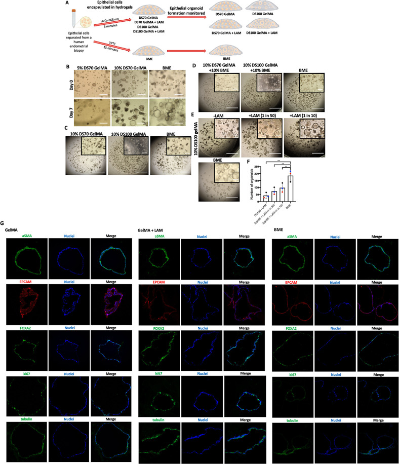
Figure 4.
Organoid formation in GelMA hydrogels is supported by stiff matrix mechanical properties and enhanced by laminin supplementation. (A) Schematic overview of experimental design. (B) Representative images of endometrial organoids in stiff (10% DS100) and soft (10% DS70) GelMA hydrogels compared to those in BME. (C) Time-lapse images of organoids passaged into either GelMA or BME after a 14-day growth period in BME. (D) Day 6 images of organoids in GelMA–BME composite hydrogels. (E) Day 6 images of organoids in DS100 GelMA hydrogels supplemented with laminin protein compared to BME. (F) Quantification of organoid forming efficiency. Each colored point represents a biological replicate (n = 3). Scale bars = 400 μm. (G) Immunofluorescent localization of alpha-smooth muscle actin (aSMA), epithelial cell adhesion molecule (EpCAM), forkhead box A2 (FOXA2), ki67, and acetylated-α-tubulin (tubulin); in day 12, organoids grown in 10% DS100 GelMA and 10% DS100 GelMA + LAM or BME hydrogels. Nuclei were visualized with Hoechst stain.

