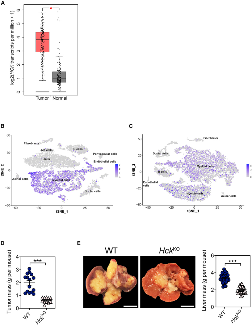
Figure 1. Genetic ablation of HCK in myeloid cells impairs PDAC tumor growth and metastasis.

(A) HCK gene expression in tumors and matched normal tissue samples of human pancreatic adenocarcinoma patients (n = 179) using the GEPIA online tool (Tang et al., 2017).
(B) tSNE plot depicting HCK gene expression in human PDAC tumors using primary data from Elyada et al. (2019).
(C) tSNE plot depicting Hck gene expression in mouse KPC PDAC tumors using primary data from Elyada et al. (2019).
(D) Mass of primary PDAC tumors from WT and HckKO hosts 5 weeks following orthotopic injection of KPC tumor cells. Each symbol represents an individual mouse. n ≥ 11 mice per group.
(E) Representative whole mounts and corresponding liver weights of WT and HckKO hosts 3 weeks after intrasplenic injection of KPC tumor cells. Scale bar: 1 cm. Each symbol represents an individual mouse. n ≥ 34 mice per group. Data represent mean ± SEM; ***p < 0.001, with statistical significance determined by an unpaired Student’s t test for comparison between two means. See also Figure S1.
