Figure 1.

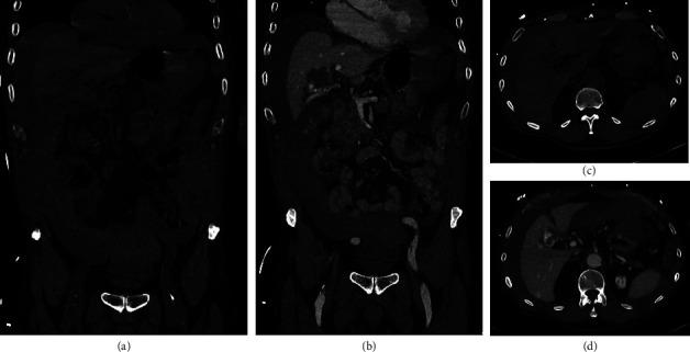
Computed tomography at admission. Computed tomography at admission is displayed. Frontal images of native (a) and venous (b) phase, as well as transverse images of native (c) and venous (d) phase, are shown.

Computed tomography at admission. Computed tomography at admission is displayed. Frontal images of native (a) and venous (b) phase, as well as transverse images of native (c) and venous (d) phase, are shown.