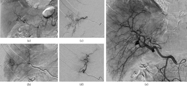
Figure 2.

Digital subtraction angiography (DSA) during the intervention. DSA images of coeliacography (a) and selective angiography of the hemangioma (b) before treatment; selective angiography during particle embolization (c) and after embolization (d), as well as final coeliacography (e) phase, is shown.
