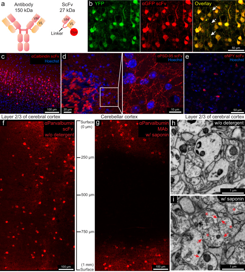
Fig. 1. Fluorescent scFv probes label brain tissues without detergents, preserving electron microscopy ultrastructure.
a Schematic of a full-length IgG antibody and an scFv probe with a fluorescent dye. b Confocal images from the cerebral cortex of a YFP-H mouse labeled with a GFP-specific scFv probe conjugated with 5-TAMRA (n = 3 experiments, all experiments mentioned refer to independent experiments). Arrows indicate unlabeled thinner neuronal processes. c Layer 2/3 of the cerebral cortex labeled with a calbindin-specific scFv probe (n = 3 experiments). d Cerebellar cortex labeled with PSD-95-specific scFv (n = 3 experiments), with an enlarged inset. e Cerebral cortex labeled with an NPY-specific scFv (n = 3 experiments). f, g Penetration depth comparison of a parvalbumin-specific scFv without detergent versus parental mAbs with 0.05% saponin (n = 2 experiments). h, i Ultrastructure comparison after 7 days incubation without detergent versus 0.05% saponin (n = 2 experiments). Arrows indicate membrane breaks; asterisks indicate abnormal vesicle-filled axonal terminals.

