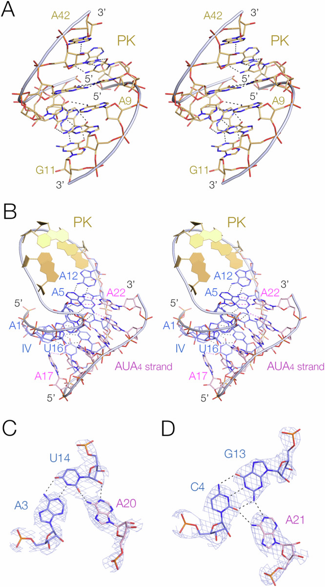
Fig. 3. The pseudoknot helix and the minor-groove triple helix in the PK hammerhead ribozyme. Parts A and B are shown in parallel-eye stereoscopic views.

A The pseudoknot helix. This comprises a 6 bp standard A-form RNA helix, with non-Watson-Crick base pairs at each end. B Helix IV, the stem of the pseudoknot helix. This forms a triple helix by binding the AUA4 sequence in its minor groove. Representative base triples in the IV triplex formed by A20 (C) and A21 (D). The electron density maps are 2Fo-Fc maps contoured at 2.5 σ.
