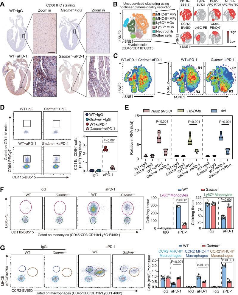
Fig. 4. Deletion of GSDME reduces aPD-1 therapy-induced myeloid cell activation and cardiac infiltration.
A Infiltration of monocytes/macrophages (MOs/MPs) into myocardium was evaluated by immunohistochemistry staining of CD68 in WT and Gsdme−/− mice received aPD-1 therapy or control IgG. The images of whole heart section and amplified specific area were illustrated. Scale bar, 200 μm. B Unsupervised clustering using t-SNE nonlinear dimensionality reduction in myeloid cells (CD45+CD11b+CD3−) cells with the CD11b, Ly6G, F4/80, MHC-II, CCR2, Ly6C and CD64 markers. The colors in the expression-level heatmaps (right panel) represent the median intensity values for each marker. C Subclusters of heart myeloid cells from WT and Gsdme−/− mice. The significantly different regions 1 to 3 (R1-3) were circled. The cells in these regions were designated in (B). D Numbers of M1-like MOs/MPs (CD11b+CD64+) in heart tissue of WT and Gsdme−/− mice were determined using flow cytometry analysis based on CD11b and CD64. E The mRNA expression of pro-inflammatory MOs/MPs-related genes (Nos2, H2-DMa and Axl) in heart tissue of WT and Gsdme−/− mice. (F) Numbers of Ly6Chigh (Ly6Chi) and Ly6Clow (Ly6Clo) monocytes in heart tissue of WT and Gsdme−/− mice were determined using flow cytometry analysis. G Numbers of CCR2−MHC-IIlo, CCR2−MHC-IIhi and CCR2+MHC-IIhi macrophages in heart tissue of WT and Gsdme−/− mice were determined using flow cytometry analysis. The data were presented as means ± SEM and analyzed by two-sided unpaired Student’s t-tests. n = 6 biologically independent experiments.

