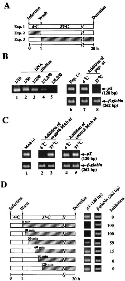
FIG. 4.
Effects of the gp21 peptide 400-429 and a neutralizing MAb against gp46 on the early stage of HTLV-1 infection. (A) Schematic representation of the experiments (Exp.) investigating HTLV-1 infection at the binding step or the postbinding step. Precooled MOLT-4 cells were inoculated with cell-free HTLV-1 at 4°C for 1 h (Exp. 2, binding step), washed, and resuspended in fresh culture medium. Then, the incubation temperature of the cells was shifted to 37°C for 20 h (Exp. 3, postbinding step). The reverse-transcribed HTLV-1 RNA in MOLT-4 cells was detected by nested PCR. (B) Inhibition of the postbinding step of HTLV-1 infection by the gp21 peptide 400-429. To detect the formation of HTLV-1 DNA, serially diluted first PCR products were used as templates for nested PCR amplification using the pXI 7341-7360 and pXI 7460-7441 primers (lanes 1 to 4). The temperatures at the time of addition of the gp21 peptide (pep.) 400-429 are indicated in panel A as hatched bars. Fiftyfold-diluted first PCR products were used as templates for nested PCR. The 120 bp of pX DNA in lanes 6 to 8 corresponds to Exp. 1 to 3 in panel A, respectively. (C) Inhibition of transmission of cell-free HTLV-1 at the binding step by anti-gp46 neutralizing MAb LAT-27. HTLV-1 was incubated with LAT-27 MAb (30 μg/ml) or control MAb against mouse recombinant IL-2 (30 μg/ml) for 1 h at 37°C prior to infection. Then, experiments were done as described above. Lane 1, no-MAb control. MAbs were added to MOLT-4 cells either at 4°C (lanes 2 and 4) or at 37°C (lanes 3 and 5). β-Globin DNA was amplified as a control. (D) Effect of the timing of the addition of the gp21 peptide 400-429 after adsorption of HTLV-1 to MOLT-4 cells on the transmission of cell-free HTLV-1. After adsorption of HTLV-1 to MOLT-4 cells at 4°C, the gp21 peptide 400-429 was added at the indicated times and the cells were cultured for 20 h in the presence of the peptide. Formation of HTLV-1 DNA in MOLT-4 cells was detected by nested PCR (120 bp of pX). β-Globin DNA was amplified as a control. Relative intensities of pX DNA bands were calculated by densitometry. Inhibition by the no-peptide control was assigned a value of 0%.

