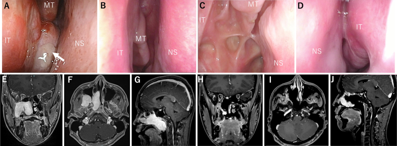
FIG. 3.
Case 7. A: Fiberscopy findings showed that the tumor (white arrow) occupied the right nasal cavity, and the posterior nostril could not be seen. BandC: Fiberscopy findings at 18 months postoperatively showed no apparent recurrence, and epithelialization was also uneventful. D: The structure of the left nasal cavity was also preserved. E–G: MRI showed that the tumor extended from the right pterygopalatine fossa to the right middle and lower nasal passages and nasopharynx. H–J: Contrast-enhanced MRI 2 years after surgery showed no evident recurrence. MT = middle turbinate.

