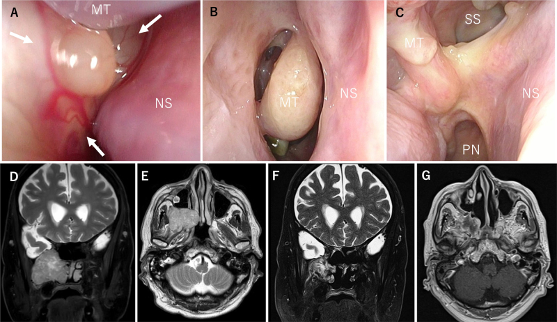
FIG. 5.
Case 17. A: Preoperative fiberscopy findings showed that the tumor (white arrows) was posterior and occupied the right nasal cavity. It took much work to observe the posterior nostril. B and C: Postoperative fiberscopy findings at 3 months showed no apparent recurrence and no complications. D and E: MRI findings showed that the tumor was centered in the pterygopalatine fossa and extended into the right nasal cavity. There was no apparent intracranial invasion. F and G: MRI 6 months postoperatively showed no apparent recurrence. PN = posterior nares; SS = sigmoid sinus.

