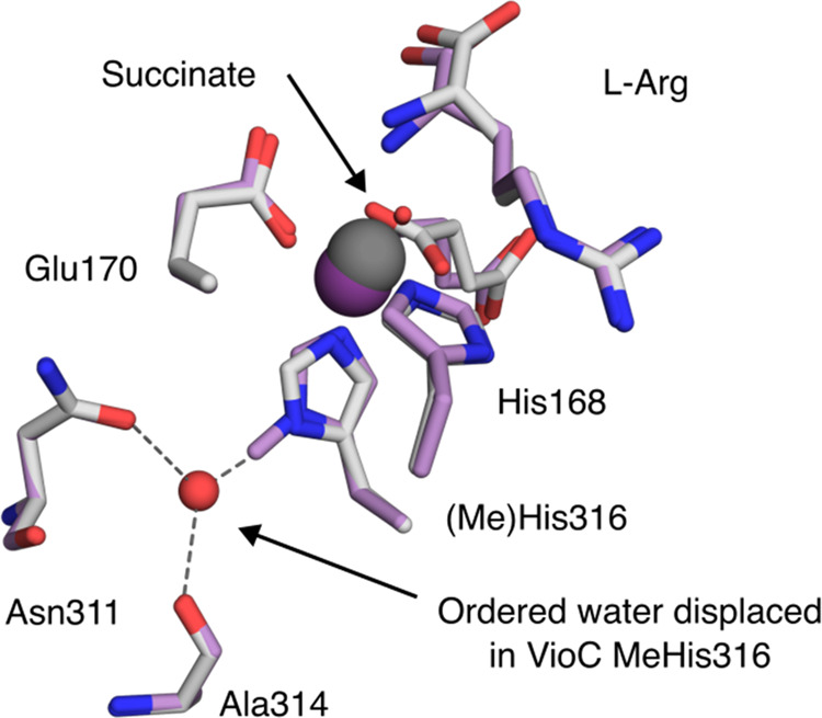
Figure 2.

Structural characterization of VioC MeHis316. The active sites of VioC (PDB: 6ALQ) and VioC MeHis316 (PDB: 9EQF). Active site residues, succinate, and bound substrate l-Arg are shown as atom-colored sticks (gray and purple carbons for VioC and VioC MeHis316, respectively). Iron is shown as dark gray or dark purple spheres in VioC (occupancy 1) and VioC MeHis316 (occupancy 0.4), respectively. An ordered water molecule that coordinates the Nδ of His316 (shown as a red sphere) is only present in the structure of VioC.
