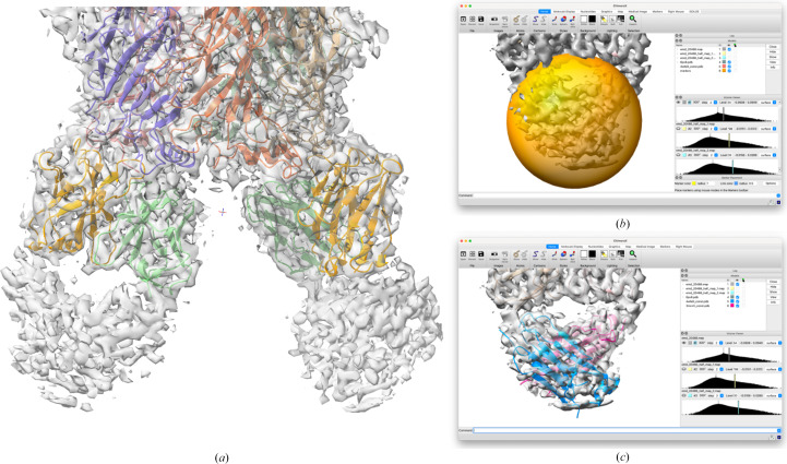
Figure 4.
Searching for the Fab constant domains in the complex with the α3β4 ganglionic nicotinic receptor (EMDB entry EMD-20488). (a) The published structure is shown in the deposited map. The modeled part of the Fab fragments consists of the variable domains of the heavy chain (light orange) and the variable domains of the light chain (light green). The map is of much poorer quality for the constant domains of these chains, at the bottom of the image. (b) The search sphere, corresponding to the uninterpreted map region in the lower right panel (a), is indicated by the yellow sphere. (c) The placed model for the constant domain from chain D of PDB entry 4wfe is shown as a blue cartoon, while the model derived from chain H of PDB entry 3mxv is shown in pink.

