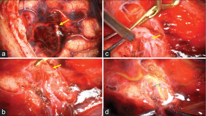
Figure 2:

Intraoperative images showing (a) a platelet plug (arrow) disrupting the arachnoid membrane corresponding to the pseudoaneurysm, (b) The disrupted middle cerebral artery with a platelet plug (arrow) after removal of the overlying arachnoid. (c) The pseudoaneurysm (arrow) was resected to a clean edge proximally and distally. (d) Demonstration of good patency throughout the anastomosis after administration of indocyanine green dye.
