
Figure 1.
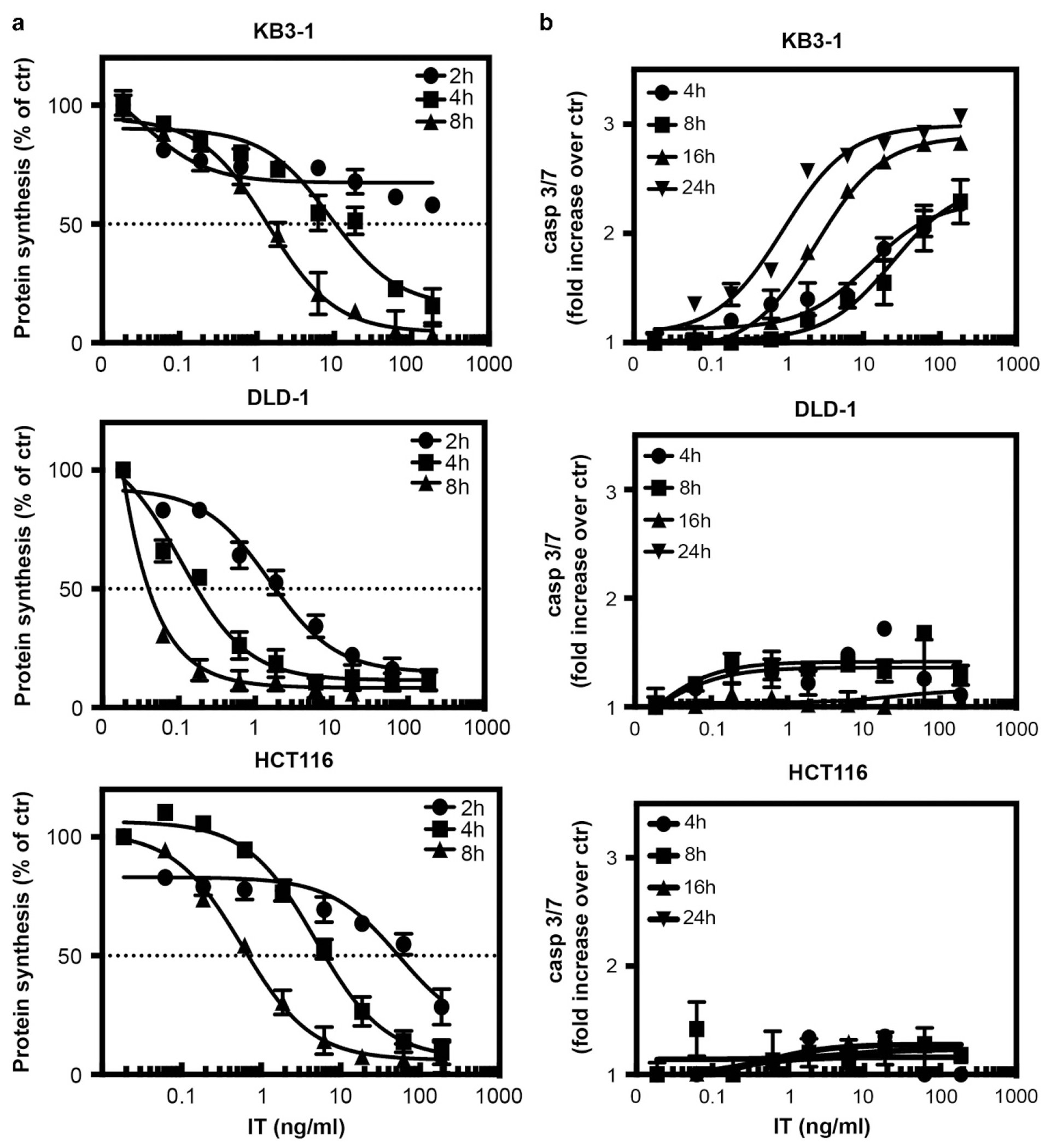
Immunotoxin-mediated inhibition of protein synthesis and activation of caspase 3/7. (a) Following the addition of various concentrations of immunotoxin to KB3-1, DLD-1 or HCT116 cells for 2, 4 or 8 h, inhibition of protein synthesis was determined by measuring the incorporation of 3H-leucine into cells. Protein synthesis was determined by measuring the incorporation of 3H-leucine into cells and the values are presented as percent compared with untreated cells. (b) KB3-1, DLD-1 and HCT116 cells were treated for 4, 8, 16 or 24 h with increasing concentrations of immunotoxin. Caspase 3/7 activity was determined using the Caspase-Glo3/7 kit. The luminescence of each well was measured and the values are presented as a fold increase relative to untreated cells. The mean values were determined from three different experiments where each point in each experiment was derived from wells in triplicate. The error bars represent the s.e.m.