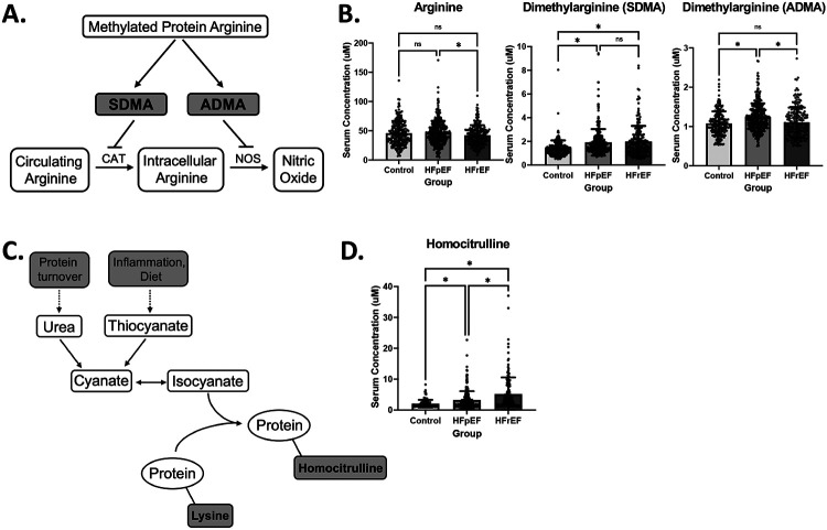
Figure 4. Asymmetric dimethylarginine is uniquely elevated in HFpEF while homocitrulline is elevated in both forms of HF.
A) Metabolic pathway depicting the formation of asymmetric dimethylarginine (ADMA) and symmetric dimethylarginine (SDMA) and subsequently their effects on arginine metabolism. B) Bar graphs depicting mean +/− SD of serum concentrations of Arginine (left), SDMA (middle), and ADMA (right) by cohort. Asterisk indicates p-value < .05. C) Metabolic pathway depicting the formation of homocitrulline residues from the incorporation of isocyanate into proteins with lysine residues. D) Bar graph depicting mean +/− SD of serum concentrations of Homocitrulline by cohort, showing significant elevation in HFrEF. Asterisk indicates p-value < .05.

