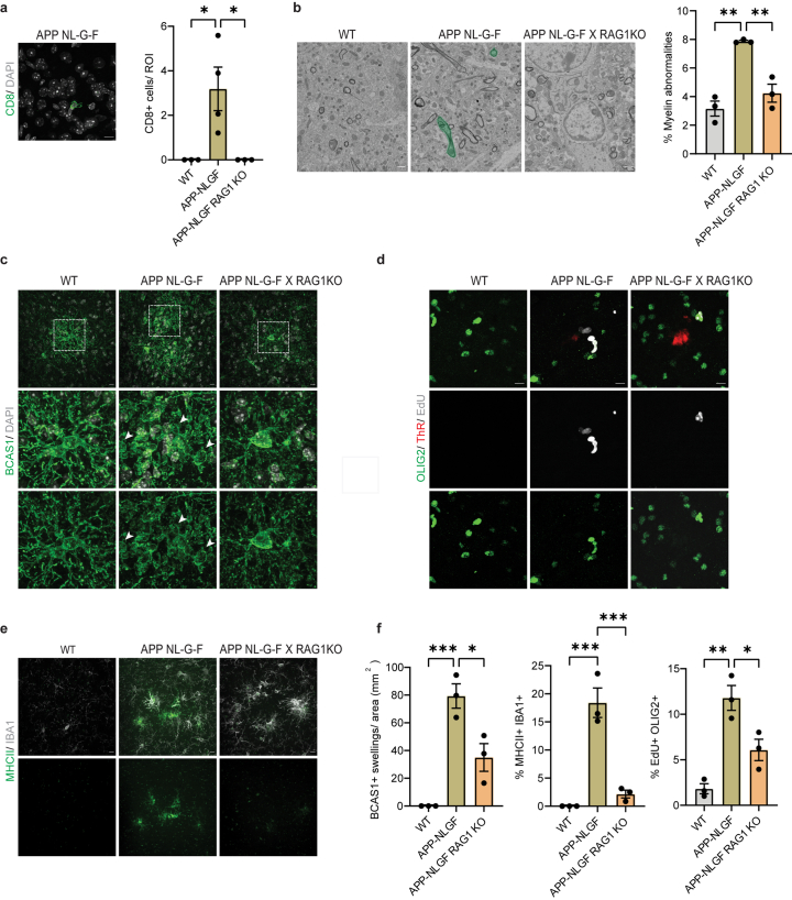
Extended Data Fig. 5. Absence of functional lymphocytes reduces oligodendrocyte and myelin damage in APP-NLGF mice.
a, Representative image showing CD8+ T cells (green) in the cortex of APP-NLGF mice. Scale bar, 10 µm. Quantifications indicate the number of CD8+ T cells in the cortex of WT, APP-NLGF and APP-NLGF X RAG1KO mice aged 12 months. Statistical significance was determined for n = 3-4 animals by one-way ANOVA with Tukey’s post hoc test (WT, APP-NLGF, *p = 0.0326; APP-NLGF, APP-NLGF X RAG1KO, *p = 0.032). b, Representative scanning electron microscopy images of the cortex of WT, APP-NLGF and APP-NLGF X RAG1KO mice aged 12 months. Myelin abnormalities are shown in green. Scale bar, 1 µm. Quantifications indicate total percentage of myelinated axons with myelin abnormalities. Statistical significance was determined for n = 3 animals by one-way ANOVA with Tukey’s post hoc test (WT, APP-NLGF, **p = 0.001; APP-NLGF, APP-NLGF X RAG1KO, **p = 0.004). c, Representative image of BCAS1+ (green) swellings lacking a nucleus (stained with DAPI) in the cortex of WT, APP-NLGF and APP-NLGF X RAG1KO mice aged 12 months. Scale bar, 10 µm. d, Representative image showing EdU+ (grey) OLIG2+ (green) cells in the cortex of WT, APP-NLGF and APP-NLGF X RAG1KO mice aged 12 months. Plaques are stained with Thiazine Red (ThR, red). Scale bar, 10 µm. e, Representative image showing MHCII+ (green) IBA1+ (grey) cells in the cortex of WT, APP-NLGF and APP-NLGF X RAG1KO mice aged 12 months. Scale bar, 10 µm. f, Quantifications indicate the number of BCAS1 swellings (WT, APP-NLGF, ***p = 0.0008; APP-NLGF, APP-NLGF X RAG1KO, *p = 0.0154), percentage of IBA1+ cells positive for MHCII (WT, APP-NLGF, ***p = 0.0004; APP-NLGF, APP-NLGF X RAG1KO, ***p = 0.0008), percentage of OLIG2+ cells also positive for EdU (WT, APP-NLGF, **p = 0.0015; APP-NLGF, APP-NLGF X RAG1KO, *p = 0.0230). Statistical significance was determined for n = 3 animals by one-way ANOVA with Tukey’s post hoc test. Each point on the graph represents one animal. Data is presented as mean ± SEM.

