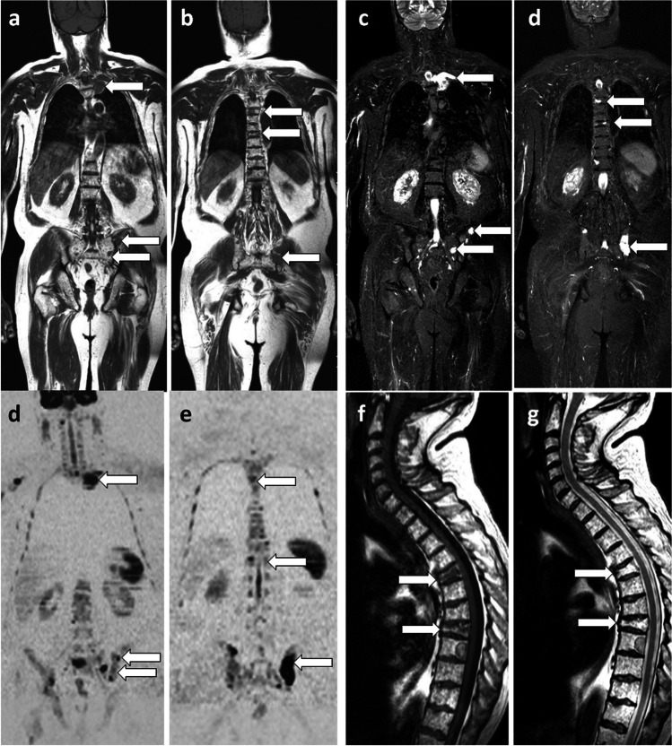
Fig. 1.
66-year-old man with newly diagnosed multiple myeloma: WB-MRI work-up. Coronal T1 (a, b), STIR (c, d) and DWI (b = 1000 s/mm2, inverted grayscale) DWI (e, f) images: multiple foci of bone marrow replacement are seen in the ribs, thoracolumbar spine and pelvis (arrows), indicating advanced disease requiring treatment. Sagittal T1 (g) and T2 (h) images of the spine: two pathologic vertebral compression fractures (arrows) are more clearly seen than on coronal sections

