Fig. 4.
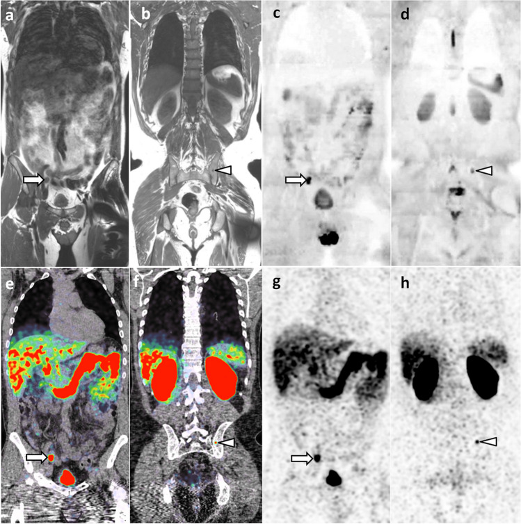
69-year-old man with newly diagnosed prostate cancer: comparison of WB-MRI and PSMA-PET/CT findings. a–d Coronal WB-MRI T1- (a, b) and high b-value (inverted grayscale window, b = 1000 s/mm2) DWI-weighted (c, d) images show right iliac lymph node metastasis (arrow) and left iliac bone metastasis (arrowhead). e–h Corresponding coronal images of PSMA-PET/CT (PET/CT fusion (e, f) and PET (g, h)) show the same lymph node (arrow) and bone (arrowhead) metastases

