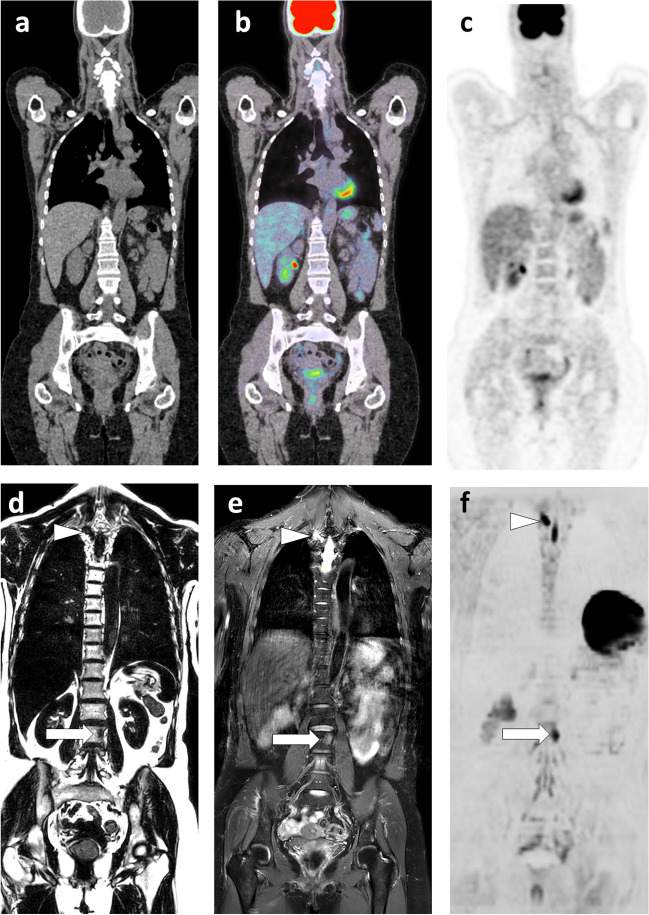
Fig. 5.
63-year-old woman with lobular carcinoma of the breast and increased tumor marker: comparison of WB-MRI and FDG-PET/CT. a–c Coronal PET/CT images (CT, fusion, PET): absence of evident lesion. d–f Corresponding coronal WB-MRI sections: fat-only (FO) (d) and water-only (WO) (f) images of a T2-weighted Dixon sequence, and coronal high b-value (inverted grayscale window, b = 1000 s/mm2) DWI image (f): bone marrow foci (metastases) at the left corporeo-pedicular junction of L3 (arrow) and the right transverse process of T3 (arrowhead). In total, three metastases were identified by this examination

